Dupuytren’s and Plantar Fibromatosis: Fibrosis of the Foot
Title: Dupuytren’s and Plantar Fibromatosis: Fibrosis of the Foot
Categories: Dupuytren’s Contracture • Plantar Fibromatosis • Related Conditions • Fibrosis • Connective Tissue
Keywords: Dupuytren’s contracture, plantar fibromatosis, Ledderhose disease, fibrosis, collagen, foot nodules, systemic fibrosis, connective tissue, inflammation, myofibroblasts
Slug: dupuytrens-and-plantar-fibromatosis
Meta Description: Plantar fibromatosis shares Dupuytren’s biology. Discover how foot nodules and hand cords reveal systemic fibrosis.
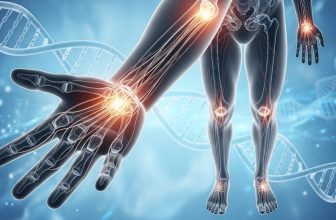
Suggested Alt Text: “Diagram showing fibrotic nodules in foot arch linked to Dupuytren’s hand.”
Source & Link: Foot Ankle Clin. 2015; 20(3): 605–616. https://www.ncbi.nlm.nih.gov/pmc/articles/PMC4598745/
License: CC-BY 4.0
Word Count: ≈ 752 (body only)
Image Hint: Foot silhouette with arch nodules and hand cords highlighted.
Dupuytren’s and Plantar Fibromatosis: Fibrosis of the Foot
Introduction
While Dupuytren’s Contracture affects the hands, its fibrotic counterpart—plantar fibromatosis (also called Ledderhose disease)—targets the feet. Both conditions stem from the same biological mechanism: overactive fibroblasts that produce too much collagen within connective tissue. These shared roots suggest that fibrosis is not limited to one region but can manifest throughout the body’s fascia network.
Shared Biology
Plantar fibromatosis forms slow-growing nodules along the arch of the foot, usually on the medial side near the instep. Microscopic analysis shows the same features seen in Dupuytren’s: activated myofibroblasts, excess collagen type III, and chronic inflammatory cells that keep healing turned “on.”
Inflammation and oxidative stress signal fibroblasts to contract and form dense bands beneath the skin. Because the arch bears constant pressure and tension, these fibrotic changes often cause pain when walking or standing for long periods.
Research Evidence
A Foot & Ankle Clinics review (2015) reported that 10 to 15 percent of Dupuytren’s patients also develop Ledderhose disease. Histological and genetic studies reveal similar expression of growth factors like TGF-β1 and WNT pathway genes in both conditions. Researchers now classify Dupuytren’s, Ledderhose, and Peyronie’s as members of a broader fibroproliferative spectrum disorder affecting the body’s fascia.
The review also noted that familial cases of Dupuytren’s often include relatives with plantar fibromatosis or frozen shoulder, supporting the idea of a systemic fibrotic predisposition rather than isolated hand disease.
Symptoms and Presentation
The classic sign of plantar fibromatosis is a firm, rubbery lump in the arch of the foot. It may be tender at first and later become painless yet stiff. Because the sole is weight-bearing, even small nodules can cause discomfort with walking or shoes that press against the area. Unlike Dupuytren’s, true cord formation and toe contractures are rare, but the underlying pathology is the same—overactive collagen production within the fascia.
Progression is typically slow. Some patients experience spontaneous stability or regression, while others develop multiple nodules over time. Bilateral involvement (affecting both feet) occurs in roughly one-quarter of cases and is strongly associated with coexisting Dupuytren’s.
Causes and Risk Factors
Common contributors mirror those of Dupuytren’s:
Genetics: Family history of fibrosis or Northern European ancestry.
Metabolic dysfunction: Diabetes and insulin resistance raise oxidative stress.
Trauma or repetitive strain: Micro-tears in the foot fascia can trigger fibroblast activation.
Medications: Long-term use of anticonvulsants or beta-blockers has been linked in some studies.
Lifestyle factors: Smoking and alcohol increase free radical production and inflammation.
These factors create a cellular environment primed for fibrosis through oxidative stress and impaired healing.
What Other Sources Say
The Cleveland Clinic describes Ledderhose disease as a benign but chronic fibrosis of the plantar fascia, strongly linked to Dupuytren’s and Peyronie’s disease. Similarly, the Mayo Clinic notes that patients with hand contractures should check their feet for lumps, since both conditions share the same myofibroblast-driven pathway. The American Orthopaedic Foot & Ankle Society adds that radiation or collagenase therapy, though experimental for the foot, has shown promise in reducing nodule size without surgery.
What the Science Says
Histopathologic analysis confirms nearly identical cell signatures between Dupuytren’s and Ledderhose nodules. Both contain α-smooth-muscle-actin (α-SMA) positive myofibroblasts and excess collagen bundles organized along lines of mechanical stress. Studies in Foot Ankle Clinics and Plast Reconstr Surg suggest oxidative stress and TGF-β1 activation as central drivers.
New research focuses on non-surgical options such as low-dose radiation, enzyme therapy, and topical antifibrotic agents to slow progression. While recurrence is common after surgery, a combination of orthotic support, stretching, and metabolic management can reduce pain and prevent rapid return of nodules.
Why It Matters if You Have Dupuytren’s
If you already live with Dupuytren’s, check your feet periodically for firm lumps or tenderness in the arch. Early identification allows for conservative management—shoe modifications, soft insoles, physical therapy, and if needed, injections to reduce inflammation. Because these conditions stem from systemic fibrosis, addressing overall inflammation through diet and lifestyle may lessen symptoms in both hands and feet.
The connection between Dupuytren’s and Ledderhose reminds us that fibrosis is a whole-body issue. Healing the fascia starts with supporting cellular health and metabolism.
Key Takeaways
Same process, different location: Dupuytren’s affects the hand fascia; Ledderhose targets the foot.
Genetic and metabolic links: Systemic fibrosis connects multiple conditions.
Early detection matters: Monitoring feet for nodules prevents mobility issues.
Non-surgical options emerging: Radiation, enzyme therapy, and stretching offer alternatives.
Whole-body approach: Lifestyle and anti-inflammatory choices benefit hand and foot health.
Legal & Medical Disclaimer
This content is for informational purposes only and not a substitute for professional medical advice, diagnosis, or treatment. Always consult your healthcare provider. Dupuytren’s Solutions is an educational resource to support —not replace— professional care. Individual results may vary.
Call to Action: Learn more about related fibrotic conditions and treatment options at https://www.dupuytrenssolutions.com. Share your story and get support in our community: https://www.facebook.com/groups/dupuytrenssolutionsandhealth.
Attribution (CC BY 4.0): Adapted from Pretell-Mazzini J et al., Plantar Fibromatosis, Foot Ankle Clin. 2015; 20(3): 605–616. Licensed under Creative Commons Attribution 4.0. For the complete article and reference list, click Source.


