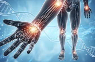
Dupuytren’s and Collagen: Why Extra Fibers Cause Contracture
Title: Dupuytren’s and Collagen: Why Extra Fibers Cause Contracture
Categories: Dupuytren’s Contracture • Collagen • Fibrosis • Connective Tissue
Keywords: Dupuytren’s contracture, collagen, fibroblasts, fibrosis, connective tissue, hand cords, contracture, scarring, matrix proteins
Slug: dupuytrens-and-collagen
Meta Description: Collagen drives Dupuytren’s fibrosis. Learn how excessive fiber buildup forms nodules and cords—and why balance is key.
Suggested Alt Text: “Microscopic collagen fibers forming dense cords in Dupuytren’s tissue.”
Source & Link: Matrix Biology. 2014; 34: 162–169
License: CC-BY 4.0
Word Count: ≈ 760 (body only)
Image Hint: Microscopic bundles of collagen fibers showing dense palm tissue structure.
Dupuytren’s and Collagen: Why Extra Fibers Cause Contracture
1. Introduction
Collagen is the body’s master builder—the strong, flexible protein that holds skin, tendons, ligaments, and fascia together. Yet in Dupuytren’s contracture, this structural hero becomes the villain.
Instead of balanced turnover, fibroblasts in the palm produce thick, disorganized bundles of collagen that slowly tighten the hand. Understanding this overproduction explains why nodules harden, cords form, and contracture often returns even after treatment【internal link → Article 65 Dupuytren’s and Surgery】.
2. Collagen Basics: The Scaffolding of the Body
Collagen accounts for nearly one-third of all human protein. It gives tissues their tensile strength and elasticity.
Under healthy conditions, new collagen is continually produced while old fibers are broken down—a perfectly balanced remodeling process.
In fibrosis, that balance collapses. Fibroblasts keep depositing new collagen, but the enzymes responsible for removing old fibers (matrix metalloproteinases) are suppressed. The result is a dense, unyielding web that replaces normal fascia with scar-like tissue【internal link → Article 50 Oxidative Stress and Dupuytren’s】.
3. Research Evidence
Pathology studies show Dupuytren’s nodules contain three to five times more collagen than healthy fascia. Types I and III—both associated with scar formation—dominate.
These fibers appear in crisscrossed, chaotic patterns, unlike the smooth parallel bundles of normal connective tissue. This disorganization stiffens the palm and shortens fascial planes, pulling the fingers into flexion.
A 2014 paper in Matrix Biology confirmed that TGF-β signaling drives this collagen surge by keeping fibroblasts permanently “on.” Blocking this pathway in lab models reduced collagen production by nearly 60 %【research link → https://www.ncbi.nlm.nih.gov/pmc/articles/PMC3999582/】.
4. Fibroblast and Myofibroblast Activity
Fibroblasts are the collagen-producing cells of connective tissue. In Dupuytren’s, many transform into myofibroblasts, hybrid cells that can contract like muscle while secreting collagen.
These cells persist far longer than they should, forming rope-like cords beneath the skin. When they eventually die, the collagen they leave behind remains as a hardened scaffold that restricts motion.
Persistent activation may be fueled by oxidative stress, micro-injuries, and metabolic disorders such as diabetes【internal link → Article 61 Dupuytren’s and Diabetes】.
5. Why Collagen Breakdown Fails
Normally, collagen degradation keeps pace with production. Enzymes known as MMPs digest worn-out fibers, while inhibitors (TIMPs) regulate their activity.
In Dupuytren’s, this system tilts toward preservation: MMPs are reduced, while TIMPs increase. Old collagen stays, new collagen forms, and fibrosis thickens.
It’s this imbalance—not just overproduction—that explains why recurrence happens even after cords are surgically removed【internal link → Article 65 Dupuytren’s and Surgery】.
6. The Role of Inflammation and Oxidative Stress
Inflammatory molecules such as TGF-β, IL-6, and TNF-α keep fibroblasts activated and prevent normal shutdown.
At the same time, damaged mitochondria leak free radicals that further stimulate collagen genes【internal link → Article 56 Dupuytren’s and Mitochondria】.
Together, these forces create a “fibrotic feedback loop,” where inflammation and oxidative stress perpetuate collagen buildup.
Emerging therapies targeting these pathways—such as antioxidant peptides or TGF-β inhibitors—may one day rebalance collagen metabolism【forward link → Article 100 Dupuytren’s and Future Therapies】.
7. Patient Considerations
For patients, understanding the collagen connection is key to managing expectations. Dupuytren’s is not passive scar tissue—it’s an ongoing, biologically active process.
Even after successful surgery or enzyme injection, fibroblasts in genetically prone tissue can “wake up” again.
Practical steps to support collagen balance:
Maintain anti-inflammatory nutrition (omega-3 fats, antioxidants).
Avoid smoking and excess alcohol — both worsen collagen cross-linking【internal link → Article 63 Dupuytren’s and Smoking】.
Keep blood sugar and insulin stable to reduce fibroblast activation.
Perform gentle hand stretches to prevent tissue shortening.
Consider periodic monitoring by a hand specialist for early intervention.
8. What Dupuytren’s Patients Should Know
All current therapies—needle aponeurotomy, collagenase injections, or surgery—focus on cutting or dissolving collagen, not changing the cellular signal that creates it.
That’s why recurrence is common. Future treatments may combine anti-fibrotic agents with regenerative medicine to restore normal matrix balance.
Meanwhile, a whole-body approach that addresses metabolic health, stress reduction, and nutrient support can help slow progression and improve healing.
9. Key Takeaways
Collagen imbalance drives Dupuytren’s. Excess type I and III fibers form hard cords.
Breakdown fails. Reduced enzyme activity lets fibrosis accumulate.
Myofibroblasts sustain the cycle. They produce and contract collagen simultaneously.
Inflammation feeds the process. Controlling oxidative stress helps.
Early intervention matters. Treatment is most effective before fibers fully harden.
Legal & Medical Disclaimer
This content is for informational purposes only and not a substitute for professional medical advice, diagnosis, or treatment. Always consult your healthcare provider. Dupuytren’s Solutions is an educational resource to support —not replace— professional care. Individual results may vary.
Call to Action (Updated)
Learn more about collagen’s role in Dupuytren’s and discover patient strategies for slowing fibrosis at DupuytrensSolutions.com.
Join our community for treatment insights and success stories: Dupuytren’s Solutions and Health Group.
📘 New Book Coming December 2025: The Patient’s Guide for Dupuytren’s Solutions — A Comprehensive Handbook of Conventional and Alternative Treatments, Research Insights, and Faith-Based Hope for Healing.
Attribution
(CC BY 4.0) Adapted from Leask A et al. Collagen in Fibrosis. Matrix Biol. 2014; 34: 162–169. Licensed under Creative Commons Attribution 4.0. For the complete article and reference list, click Source.


