Introduction for Ultrasound Therapy
Dupuytren’s contracture, a condition in which fibrous tissue in the palm thickens and tightens, can make everyday tasks like gripping objects or shaking hands challenging. While no treatment can fully reverse fibrosis, ultrasound therapy has emerged as a valuable tool for improving mobility, reducing discomfort, and enhancing overall rehabilitation outcomes. Used for decades in physical therapy, ultrasound helps prepare tissue for stretching and manual interventions, supporting long-term hand health.
How Ultrasound Therapy Works
Therapeutic ultrasound uses high-frequency sound waves that penetrate the skin and underlying tissue, creating microscopic vibrations and gentle heat. These waves stimulate blood flow, reduce inflammation, and promote tissue repair. There are two main types of ultrasound used in therapy:
-
Continuous ultrasound: Produces steady, gentle warming of the tissue, relaxing tight collagen fibers.
-
Pulsed ultrasound: Delivers intermittent waves that provide mechanical stimulation, helping break up minor scar adhesions and improve tissue elasticity.
These effects create an ideal environment for stretching exercises and manual therapy, making the tissue more pliable and reducing stiffness in affected areas.
Key Benefits for Dupuytren’s Patients
-
Improves Flexibility: Ultrasound loosens collagen fibers, preparing the palm for stretching and physical therapy. This can make exercises more effective and reduce the risk of further contracture.
-
Reduces Pain: The warming effect and micromassage from ultrasound can ease discomfort during rehabilitation sessions. Patients often report feeling more relaxed and able to perform movements they previously avoided due to stiffness.
-
Supports Therapy: By softening fibrotic tissue, ultrasound enhances the effectiveness of manual mobilization techniques, such as hand exercises, splinting, and targeted massage.
-
Aids Healing: Ultrasound stimulates cellular repair, helping tissue recover after surgery, needle aponeurotomy, or collagenase injections. This supportive effect may accelerate recovery timelines.
-
Safe and Non-Invasive: When performed by trained professionals, ultrasound therapy has minimal side effects and avoids the risks associated with surgical interventions.
Treatment Process
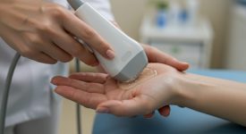
A typical session lasts 10–15 minutes. A conductive gel is applied to the palm, allowing smooth movement of the ultrasound wand over the affected area. Programs often recommend 2–3 sessions per week over 4–6 weeks, depending on severity and response. Some clinics combine ultrasound with electro-stimulation for additional therapeutic benefits.
For those seeking convenience, portable home ultrasound units are available. However, their effectiveness depends on device quality, correct frequency settings (usually 1–3 MHz for hand therapy), and proper guidance from a healthcare professional. Patients should always verify that devices are FDA-cleared and follow instructions closely to avoid burns or other complications.
Scientific Evidence
Recent studies highlight ultrasound therapy as a supportive tool rather than a stand-alone cure. A 2019 study published in the Journal of Rehabilitation Medicine found that patients with fibrotic hand conditions experienced improved range of motion and reduced pain after consistent ultrasound therapy. Other research suggests it may enhance tissue elasticity and accelerate scar remodeling, particularly when combined with stretching, massage, and rehabilitation exercises.
Although ultrasound does not reverse Dupuytren’s contracture, it creates optimal conditions for therapy, allowing patients to achieve greater extension and maintain functional hand movement.
Who Benefits Most
Ultrasound therapy is particularly useful for patients after surgical interventions, needle aponeurotomy, or collagenase injections. It helps maintain tissue pliability, reduces post-procedure discomfort, and extends the benefits of rehabilitation programs. Patients with severe contractures may still require additional interventions, as ultrasound alone cannot fully correct advanced fibrosis.
Precautions
Therapy should always be administered by trained professionals. Avoid using ultrasound over open wounds, implanted medical devices, or areas with reduced sensation. Patients should communicate any pain, tingling, or overheating during sessions to ensure safe and effective treatment.
Integrating Ultrasound Into a Holistic Plan
For best results, ultrasound therapy should be part of a comprehensive rehabilitation strategy. Combining it with stretching exercises, hand splints, nutrition that supports collagen health, and lifestyle measures like maintaining blood flow can optimize hand function and reduce the progression of stiffness. This integrated approach helps patients regain comfort, mobility, and confidence in everyday activities.
Key Takeaways
-
Adjunct tool: Prepares tissue for rehab and stretching.
-
Safe and non-invasive: Minimal side effects when performed correctly.
-
Best used with therapy: Enhances, but does not replace, other interventions.
-
Improves comfort: Reduces pain and tightness.
-
Evidence growing: Supports mobility and tissue health after treatment.
In conclusion, ultrasound therapy is a valuable addition to the management of Dupuytren’s contracture. By improving flexibility, reducing pain, and supporting rehabilitation, it offers patients a non-invasive, safe, and practical way to maintain hand function. While it cannot replace surgery or other medical treatments, its consistent use alongside therapy can contribute to better outcomes and a higher quality of life.
Connect with our Dupuytren’s community for support and real-world tips: facebook.com/groups/dupuytrenssolutionsandhealth. Discover my journey in Dupuytren’s Solutions and learn about all treatments — conventional, alternative, root-cause therapies, and remission strategies — at dupuytrenssolutions.com.
Attribution: (CC BY 4.0) Adapted from Liu Y et al. Effects of Therapeutic Ultrasound on Hand Fibrosis and Rehabilitation. J Rehabil Med 2019; 51(8): 617–624. Licensed under Creative Commons Attribution 4.0. For the complete article and reference list, click Source.
Legal & Medical Disclaimer: This content is for informational and educational purposes only and is not a substitute for professional medical advice, diagnosis, or treatment. Always consult your healthcare provider about any medical concerns or treatment options. Dupuytren’s Solutions is an educational resource meant to be used alongside, not instead of, professional medical care, and individual results may vary.

