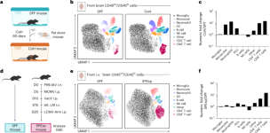
Physiological microbial exposure normalizes memory T cell surveillance of the brain and modifies host seizure outcomes
Svenningsson, A., Andersen, O., Edsbagge, M. & Stemme, S. Lymphocyte phenotype and subset distribution in normal cerebrospinal fluid. J. Neuroimmunol. 63, 39–46 (1995).
Smolders, J. et al. Tissue-resident memory T cells populate the human brain. Nat. Commun. 9, 4593 (2018).
Pappalardo, J. L. et al. Transcriptomic and clonal characterization of T cells in the human central nervous system. Sci. Immunol. 5, eabb8786 (2020).
Piehl, N. et al. Cerebrospinal fluid immune dysregulation during healthy brain aging and cognitive impairment. Cell 185, 5028–5039 (2022).
Smolders, J. et al. Characteristics of differentiated CD8+ and CD4+ T cells present in the human brain. Acta Neuropathol. 126, 525–535 (2013).
Mix, M. R. & Harty, J. T. Keeping T cell memories in mind. Trends Immunol. 43, 1018–1031 (2022).
Ayasoufi, K. et al. Brain resident memory T cells rapidly expand and initiate neuroinflammatory responses following CNS viral infection. Brain Behav. Immun. 112, 51–76 (2023).
Pasciuto, E. et al. Microglia require CD4 T cells to complete the fetal-to-adult transition. Cell 182, 625–640 (2020).
Wakim, L. M., Woodward-Davis, A. & Bevan, M. J. Memory T cells persisting within the brain after local infection show functional adaptations to their tissue of residence. Proc. Natl Acad. Sci. USA 107, 17872–17879 (2010).
Wakim, L. M. et al. The molecular signature of tissue resident memory CD8 T cells isolated from the brain. J. Immunol. 189, 3462–3471 (2012).
Landrith, T. A. et al. CD103+ CD8 T cells in the Toxoplasma-infected brain exhibit a tissue-resident memory transcriptional profile. Front. Immunol. 8, 335 (2017).
Garber, C. et al. T cells promote microglia-mediated synaptic elimination and cognitive dysfunction during recovery from neuropathogenic flaviviruses. Nat. Neurosci. 22, 1276–1288 (2019).
Vincenti, I. et al. Tissue-resident memory CD8+ T cells cooperate with CD4+ T cells to drive compartmentalized immunopathology in the CNS. Sci. Transl. Med. 14, eabl6058 (2022).
Fransen, N. L. et al. Tissue-resident memory T cells invade the brain parenchyma in multiple sclerosis white matter lesions. Brain 143, 1714–1730 (2020).
Bernard-Valnet, R. et al. Influenza vaccination induces autoimmunity against orexinergic neurons in a mouse model for narcolepsy. Brain 145, 2018–2030 (2022).
Urban, S. L. et al. Peripherally induced brain tissue-resident memory CD8+ T cells mediate protection against CNS infection. Nat. Immunol. 21, 938–949 (2020).
Dulken, B. W. et al. Single-cell analysis reveals T cell infiltration in old neurogenic niches. Nature 571, 205–210 (2019).
Cassidy, B. R., Sonntag, W. E., Leenen, P. J. M. & Drevets, D. A. Systemic Listeria monocytogenes infection in aged mice induces long-term neuroinflammation: the role of miR-155. Immun. Ageing 19, 25 (2022).
Wang, X. et al. CD8+ T cells exacerbate AD-like symptoms in mouse model of amyloidosis. Brain Behav. Immun. 122, 444–455 (2024).
Masopust, D. & Soerens, A. G. Tissue-resident T cells and other resident leukocytes. Annu. Rev. Immunol. 37, 521–546 (2019).
Martin, M. D. & Badovinac, V. P. Defining memory CD8 T cell. Front. Immunol. 9, 2692 (2018).
Frieser, D. et al. Tissue-resident CD8+ T cells drive compartmentalized and chronic autoimmune damage against CNS neurons. Sci. Transl. Med. 14, eabl6157 (2022).
Chen, X. et al. Microglia-mediated T cell infiltration drives neurodegeneration in tauopathy. Nature 615, 668–677 (2023).
Ning, J. et al. Functional virus-specific memory T cells survey glioblastoma. Cancer Immunol. Immunother. 71, 1863–1875 (2022).
Ghazanfari, N. et al. CD8+ and CD4+ T cells infiltrate into the brain during Plasmodium berghei ANKA infection and form long-term resident memory. J. Immunol. 207, 1578–1590 (2021).
Casey, K. A. et al. Antigen-independent differentiation and maintenance of effector-like resident memory T cells in tissues. J. Immunol. 188, 4866–4875 (2012).
Mix, M. R. et al. Repetitive antigen stimulation in the periphery dictates the composition and recall responses of brain-resident memory CD8+ T cells. Cell Rep. 44, 115247 (2025).
Gate, D. et al. Clonally expanded CD8 T cells patrol the cerebrospinal fluid in Alzheimer’s disease. Nature 577, 399–404 (2020).
Chang, J. W. et al. Clonally focused public and private T cells in resected brain tissue from surgeries to treat children with intractable seizures. Front. Immunol. 12, 664344 (2021).
Hamilton, S. E. et al. New insights into the immune system using dirty mice. J. Immunol. 205, 3–11 (2020).
Rehermann, B., Graham, A.L., Masopust, D. & Hamilton, S.E. Integrating natural commensals and pathogens into preclinical mouse models. Nat. Rev. Immunol. 25, 385–397 (2024).
Bruno, P., Schüler, T. & Rosshart, S.P. Born to be wild: utilizing natural microbiota for reliable biomedical research. Trends Immunol. 46, 17–28 (2025).
Beura, L. K. et al. Normalizing the environment recapitulates adult human immune traits in laboratory mice. Nature 532, 512–516 (2016).
Reese, T. A. et al. Sequential infection with common pathogens promotes human-like immune gene expression and altered vaccine response. Cell Host Microbe 19, 713–719 (2016).
Rosshart, S. P. et al. Laboratory mice born to wild mice have natural microbiota and model human immune responses. Science 365, eaaw4361 (2019).
Huggins, M. A. et al. Microbial exposure enhances immunity to pathogens recognized by TLR2 but increases susceptibility to cytokine storm through TLR4 sensitization. Cell Rep. 28, 1729–1743 (2019).
Fiege, J. K. et al. Mice with diverse microbial exposure histories as a model for preclinical vaccine testing. Cell Host Microbe 29, 1815–1827 (2021).
Block, K. E. et al. Physiological microbial exposure transiently inhibits mouse lung ILC2 responses to allergens. Nat. Immunol. 23, 1703–1713 (2022).
Berton, R. R., Jensen, I. J., Harty, J. T., Griffith, T. S. & Badovinac, V. P. Inflammation controls susceptibility of immune-experienced mice to sepsis. Immunohorizons 6, 528–542 (2022).
Sjaastad, F. V. et al. Reduced T cell priming in microbially experienced ‘dirty’ mice results from limited IL-27 production by XCR1+ dendritic cells. J. Immunol. 209, 2149–2159 (2022).
Burger, S. et al. Natural microbial exposure from the earliest natural time point enhances immune development by expanding immune cell progenitors and mature immune cells. J. Immunol. 210, 1740–1751 (2023).
Martin, M. D. et al. CD115+ monocytes protect microbially experienced mice against E. coli-induced sepsis. Cell Rep. 42, 113345 (2023).
Li, Y. et al. Sequential early-life viral infections modulate the microbiota and adaptive immune responses to systemic and mucosal vaccination. PLoS Pathog. 20, e1012557 (2024).
Anderson, K. G. et al. Intravascular staining for discrimination of vascular and tissue leukocytes. Nat. Protoc. 9, 209–222 (2014).
DuPage, M. & Bluestone, J. A. Harnessing the plasticity of CD4+ T cells to treat immune-mediated disease. Nat. Rev. Immunol. 16, 149–163 (2016).
Wang, H., Gavil, N. V., Koewler, N., Masopust, D. & Jameson, S. C. Parabiosis in mice to study tissue residency of immune cells. Curr. Protoc. 2, e446 (2022).
Hayward, S. L. et al. Environmental cues regulate epigenetic reprogramming of airway-resident memory CD8+ T cells. Nat. Immunol. 21, 309–320 (2020).
Watanabe, R. et al. Human skin is protected by four functionally and phenotypically discrete populations of resident and recirculating memory T cells. Sci. Transl. Med. 7, 279ra239 (2015).
Bartolome-Casado, R. et al. Resident memory CD8 T cells persist for years in human small intestine. J. Exp. Med. 216, 2412–2426 (2019).
Christo, S. N. et al. Discrete tissue microenvironments instruct diversity in resident memory T cell function and plasticity. Nat. Immunol. 22, 1140–1151 (2021).
Fonseca, R. et al. RUNX3 drives a CD8+ T cell tissue residency program that is absent in CD4+ T cells. Nat. Immunol. 23, 1236–1245 (2022).
Dani, N. et al. A cellular and spatial map of the choroid plexus across brain ventricles and ages. Cell 184, 3056–3074 (2021).
Constant, O. et al. Role of dendritic cells in viral brain infections. Front. Immunol. 13, 862053 (2022).
Manglani, M. & McGavern, D. B. Intravital imaging of neuroimmune interactions through a thinned skull. Curr. Protoc. Immunol. 120, 24.2.1–24.2.12 (2018).
GBD 2016 Neurology Collaborators.Global, regional, and national burden of neurological disorders, 1990–2016: a systematic analysis for the Global Burden of Disease Study 2016. Lancet Neurol. 18, 459–480 (2019).
GBD 2021 Nervous System Disorders Collaborators. Global, regional, and national burden of disorders affecting the nervous system, 1990–2021: a systematic analysis for the Global Burden of Disease Study 2021. Lancet Neurol. 23, 344–381 (2024).
Grone, B. P. & Baraban, S. C. Animal models in epilepsy research: legacies and new directions. Nat. Neurosci. 18, 339–343 (2015).
Buchanan, G. F., Murray, N. M., Hajek, M. A. & Richerson, G. B. Serotonin neurones have anti-convulsant effects and reduce seizure-induced mortality. J. Physiol. 592, 4395–4410 (2014).
Rogawski, M. A. Molecular targets versus models for new antiepileptic drug discovery. Epilepsy Res. 68, 22–28 (2006).
Toman, J. E., Swinyard, E. A. & Goodman, L. S. Properties of maximal seizures, and their alteration by anticonvulant drugs and other agents. J. Neurophysiol. 9, 231–239 (1946).
Narasimhan, H. et al. An aberrant immune–epithelial progenitor niche drives viral lung sequelae. Nature 634, 961–969 (2024).
Evrard, M. et al. Single-cell protein expression profiling resolves circulating and resident memory T cell diversity across tissues and infection contexts. Immunity 56, 1664–1680 (2023).
Su, W. et al. CXCR6 orchestrates brain CD8+ T cell residency and limits mouse Alzheimer’s disease pathology. Nat. Immunol. 24, 1735–1747 (2023).
Leitner, D. et al. Similar brain proteomic signatures in Alzheimer’s disease and epilepsy. Acta Neuropathol. 147, 27 (2024).
Kumar, P. et al. Single-cell transcriptomics and surface epitope detection in human brain epileptic lesions identifies pro-inflammatory signaling. Nat. Neurosci. 25, 956–966 (2022).
Gate, D. et al. CD4+ T cells contribute to neurodegeneration in Lewy body dementia. Science 374, 868–874 (2021).
Galiano-Landeira, J., Torra, A., Vila, M. & Bove, J. CD8 T cell nigral infiltration precedes synucleinopathy in early stages of Parkinson’s disease. Brain 143, 3717–3733 (2020).
Kedia, S. et al. T cell-mediated microglial activation triggers myelin pathology in a mouse model of amyloidosis. Nat. Neurosci. 27, 1468–1474 (2024).
Sun, E.D. et al. Spatial transcriptomic clocks reveal cell proximity effects in brain ageing. Nature 638, 160–171 (2024).
Badovinac, V. P., Messingham, K. A., Jabbari, A., Haring, J. S. & Harty, J. T. Accelerated CD8+ T-cell memory and prime-boost response after dendritic-cell vaccination. Nat. Med. 11, 748–756 (2005).
Van Braeckel-Budimir, N., Varga, S. M., Badovinac, V. P. & Harty, J. T. Repeated antigen exposure extends the durability of influenza-specific lung-resident memory CD8+ T cells and heterosubtypic immunity. Cell Rep. 24, 3374–3382 (2018).
Anthony, S. M. et al. Protective function and durability of mouse lymph node-resident memory CD8+ T cells. eLife 10, e68662 (2021).
Hickman, H. D. Imaging CD8+ T cells during diverse viral infections. Intravital 4, e1055425 (2015).


